Posts Tagged ‘protein electrophoresis’

What You Don’t See Truly Matters

 :: Posted by American Biotechnologist on 11-13-2013

All gels are not created equal when it comes to protein transfer. Mini-PROTEAN® TGX™ precast gels, together with the Trans-Blot® Turbo™ transfer system, offer you a complete solution for achieving fast electrophoresis and transfer times. In addition to speed, these innovative gels deliver maximum transfer of proteins onto membranes so you don’t see proteins left behind on the gel, ensuring quality western blotting results you can count on every time.

For more information visit Bio-Rad Laboratories TGX™ precast gel page.

Chocolate Cake Electrophoresis

 :: Posted by American Biotechnologist on 05-30-2012

Checkout how students from the University of California, Davis demystify the process of electrophoresis using everyone’s favorite comfort food in an intriguing analogy.

Please note that the American Biotechnologist is not responsible for the accuracy of third party YouTube videos. If you have a comment on the video, please write it in the comment section of this post so that it will generate discussion among our scientific colleagues.

How to make an SDS-PAGE gel

 :: Posted by American Biotechnologist on 03-22-2012

Here’s a great tutorial that we found on YouTube showing viewers how to cast an SDS-PAGE gel. The video is very useful for training new students and a good refresher for others who haven’t run a PAGE gel in a while (if those types of people really exist…shame on you!). Please note that this is an independently created video (thanks labtricks and we would love to hear (and publicize) other independently created videos that you’ve found to be useful (with full credit of course).

Transfering high molecular weight proteins

 :: Posted by American Biotechnologist on 02-20-2012

We are all intimately familiar with protein blotting techniques which have been a cornerstone of the biochemicstry/biology lab for the past 30 years.

As is well known, the efficiency of protein migration is affected by various factors including the size and charge of the protein, and protocol optimization is often needed on a protein-specific basis. In fact, it can be particularly challenging to transfer large molecular weight proteins alongside small molecular weight proteins, as transfer conditions may cause small proteins to blow through the membrane.

Currently there are three popular techniques for protein transfer: the tank transfer, the semi-dry blotting method and the fast blotting “turbo” technique (for transfer within 3-10 minutes).

In the attached paper, Transfer of high molecular weight proteins to membranes: a comparison of transfer efficiency between blotting systems, Bio-Rad Laboratories presents a comparison of the various blotting techniques across a wide range of molecular weights with a particular emphasis on large proteins (more than 200kD).

A primer on fluorescence detection

 :: Posted by American Biotechnologist on 01-31-2012

Yesterday we told you about how to get more data from your western blots by utilizing multiplex fluorescent detection. Today, we will provide you with a primer on fluorescent detection taken from the Bio-Rad Laboratories Protein Blotting Guide.

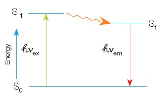
In fluorescence, a high-energy photon (ℎVex) excites a fluorophore, causing it to leave the ground state (S0) and enter a higher energy state (S’1). Some of this energy dissipates, allowing the fluorophore to enter a relaxed excited state (S1). A photon of light is emitted (ℎVem), returning the fluorophore to the ground state. The emitted photon is of a lower energy
(longer wavelength) due to the dissipation of energy while in the excited state.

When using fluorescence detection, consider the following optical characteristics of the fluorophores to optimize the signal:

  • Quantum yield — efficiency of photon emission after absorption of a photon. Processes that return the fluorophore to the ground state but do not result in the emission of a fluorescence photon lower the quantum yield.Fluorop hores with higher quantum yields are generally brighter
  •  

  • Extinction coefficient — measure of how well a fluorophore absorbs light at a specific wavelength. Since absorbance depends on path length and concentration (Beer’s Law), the extinction coefficient is usually expressed in cm–1 M–1. As with quantum yield, fluorophores with higher extinction coefficients are usually brighter
  •  

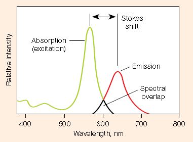
  • Stokes shift — difference in the maximum excitation and emission wavelengths of a fluorophore. Since some energy is dissipated while the fluorophore is in the excited state, emitted photons are of lower energy (longer wavelength) than the light used for excitation. Larger Stokes shifts minimize overlap between the excitation and emission wavelengths, increasing the detected signal
  •  

  • Excitation and emission spectra — excitation spectra are plots of the fluorescence intensity of a fluorophore over the range of excitation wavelengths; emission spectra show the emission wavelengths of the fluorescing molecule. Choose fluorophores that can be excited by the light source in the imager and that have emission spectra that can be captured by the instrument. When performing multiplex western blots, choose fluorophores with minimally overlapping spectra to avoid channel crosstalk
  • For more information be sure to download the Protein Blotting Guide from Bio-Rad Laboratories.